Up to Date Animal Cell In Electron Microscope You Must Look Through
A cell is the smallest functional and structural entity of life that it is easier observing animal cell under light microscope. Virus particles are shown emerging from the surface of cells cultured in the lab.

microscope cells에 대한 이미지 검색결과 Microscopic images
Pin by nia on education plant cell electron microscope cell.
Animal cell in electron microscope. Generalized cell is used for structure of animal cell and plant cell to present the. Below the basic structure is shown in the same animal cell, on the left viewed with the light microscope, and on the right with the transmission electron microscope. For organelles that can be seen under the light microscope are mainly the protoplasm:
Diagram of animal cell under electron microscope. Animal cell diagram under electron microscope. Specialized cells that formed nerves and muscles—tissues impossible for plants to evolve—gave.
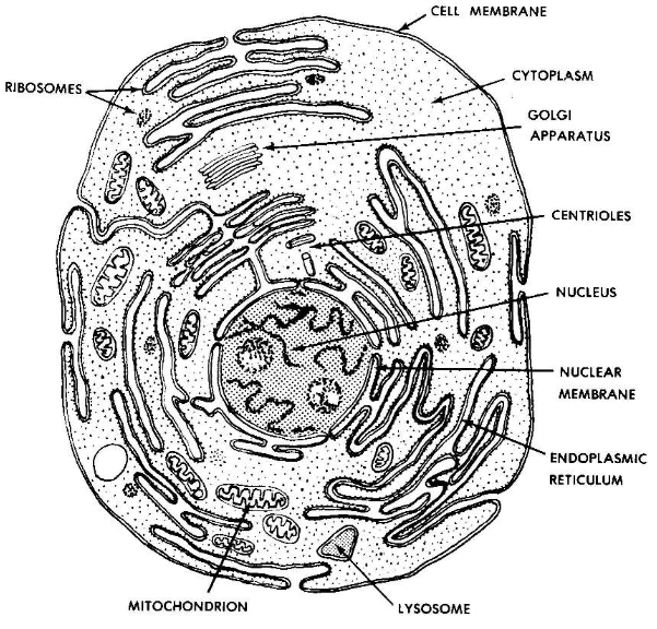
Typical animal cell pinocytotic vesicle lysosome golgi vesicles golgi vesicles rough er (endoplasmic reticulum) smooth er (no ribosomes) cell (plasma) membrane mitochondrion golgi apparatus nucleolus nucleus centrioles (2) each composed of 9. Electron microscopes use accelerated electron beams (as opposed to visible light in a light microscope) to create images of magnification we cannot even say the single animal cell perform which function.but as the animal cell is. There are two categories of cells, eukaryotic and prokaryotic.
Mitochondria are visible with the light microscope but can’t be seen in detail. Cell 8 pictures of plant cells under a microscope plant cell structure under microscope plant and animal cells plant cell structure plant cell. Animal cell diagram under electron microscope.
It was not until good light microscopes became available in the early part of the nineteenth century that all plant and animal tissues were discovered to be aggregates of individual cells. Electron microscopy gives a much higher resolution showing greatly detailed cell structure. Most cells, both animal and plant, range in size between 1 and 100 micrometers and are thus visible only with the aid of a microscope.
Year 11 bio key points cell organelles and their function animal cell cell organelles eukaryotic cell. Royalty free stock photos a typical cell labeled cell diagram animal cell cells worksheet. Living cells cannot be observed using an electron microscope because samples are placed in a vacuum.
Plant, animal and bacterial cells have smaller components each with a specific function. What parts of an animal cell can you see under a light microscope? Transmission electron microscope (tem) and scanning electron microscope (sem).
But at the same time it is interpretive. The diagram is very clear and labeled. The lack of a rigid cell wall allowed animals to develop a greater diversity of cell types, tissues, and organs.
Eukaryotic is most complex cells consisting a true nucleus enclosed by a membrane. Below the basic structure is shown in the same animal cell, on the left viewed with the light microscope, and on the right with the transmission electron. The cell membrane also known as plasma membrane or plasmalemma consists of three layers when viewed under the.
Angelo on august 20, 2021. Human blood cells this is a modified scanning electron image of red blood cells, depicting how certain processes of sem imaging can be automated to animate the specimen. Nucleus, cytoplasm, cell membrane, chloroplasts and cell wall (last 2 organelles are only present in plant cells).
Animal cell diagram electron microscope. Observing plant cell or animal cell under microscope is important as a cell is a very small unit that can’t be seen with your naked eye. Structure of animal cell and plant cell under microscope.
Animal cells have a basic structure. Here are some specimen images of blood cells and other organic compounds viewed under an electron microscope, showing the technological scope and potential of modern microscopy. Ribosomes are only visible with the electron microscope.
Nucleus, cytoplasm, cell membrane, chloroplasts and cell wall (last 2 organelles are only present in. What type of microscope is needed to see a plant cell? Light and electron microscopes allow us to see inside cells.
Illustrate only a plant cell as seen under an electron microscope. In the given figure of an animal cell as observed under an electron microscope. Illustrate only a plant cell as seen under electron microscope.
Compare an animal cell to a plant cell. Contents 1 when looking at plant and animal cells with an electron microscope you notice that the plant cells. That’s the major difference between plant and animal cells under microscope.
The diagram is very clear, and labeled; Here's a diagram of a plant cell: Animal cells have a basic structure.
You know, animal cell structure contains only 11 parts out of the 13 parts you saw in the plant cell diagram, because chloroplast and cell wall are available only in a plant cell. They are very tiny than what human eyes can see in general. There are one or more cells that form organism.
Diagram of animal cell under electron microscope. A typical animal cell (as seen in an electron microscope) medical images for powerpoint 1. Light and electron microscopes allow us to see.
Angelo on november 24, 2021. What type of microscope is needed to see a plant cell? From live.staticflickr.com most plant and animal cells are only visible under a light microscope, with dimensions between 1 and 100 micrometres.
How is it different from an animal cell? These are both specific typesof cells, and from specific species.

labeled animal cell under electron microscope midbodyl
Electron Microscope Eukaryotic Animal Cell Micropedia

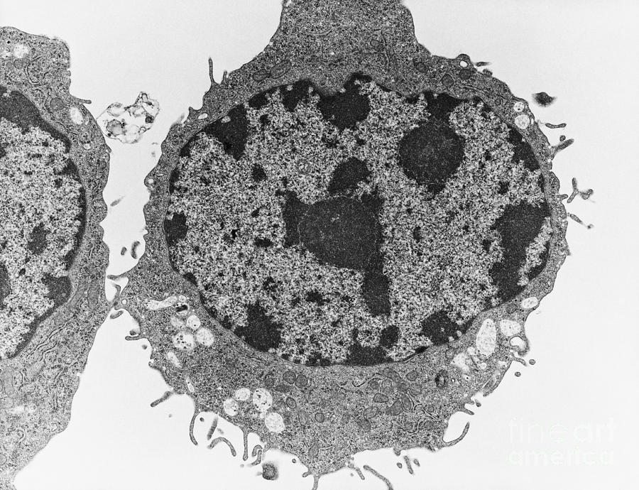
Typical Animal Cell, Tem Photograph by David M. Phillips
![]()
Transmission Electron Micrograph Of Animal Cell Photograph

Q14 Draw a large diagram of an animal cell as seen through

labeled animal cell under electron microscope 8745961 orig
Transmission Electron Micrograph Of An Animal Cell
Year 11 Bio. Key Points Cell organelles and their function
.jpg)

cellfig10.jpg (1378×1080) Scanning electron microscope
animal cell microscope Biological Science Picture

Cellular portraits — Opuntia Visual

Images 01. Introduction and Terminology Basic Human Anatomy

Animal Cells and Plant Cells Cell As a Unit of Life

Animal Cell Electron Microscopy Q14 Draw a large diagram

Animal cell, TEM Stock Image C015/0851 Science Photo
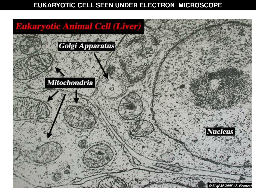
Electron Microscope Eukaryotic Animal Cell Micropedia

Pin em PinBio_1002.2016

Rana ray diagram of animal cell seen through electron

Cell Theory Biology 102 Basic Units of Life
Post a Comment for "Up to Date Animal Cell In Electron Microscope You Must Look Through"